Scienze e Natura

Twitter cambia logo e linguaggio: tutte le novità annunciate da Elon Musk
Twitter cambia logo: al posto dell’uccellino che cinguetta ci sarà una X su sfondo nero. Il link nella presentazione è già cambiato in X.com. È …
Leggi tutto
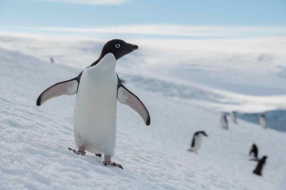
Come fermare il surriscaldamento globale? I consigli dell’esperta
Emanuela Piervitali, responsabile della Sezione climatologia operativa dell’ISPRA (Istituto Superiore per la Protezione e la ricerca ambientale): “Ci sono modi per contrastare l’aumento delle temperature” Il …
Leggi tutto
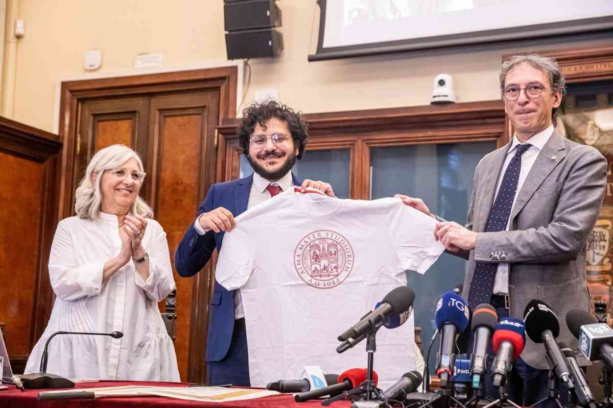
Agcom, nuove regole per gli influencer: “Devono rispettare le regole sull’audiovisivo”
Cambieranno le regole per Youtuber e distributori di materiali digitali: “Saranno soggetti alla stessa regolamentazione prevista dal Testo Unico” Cambiano le regole per gli influencer. …
Leggi tuttoSalute

Green
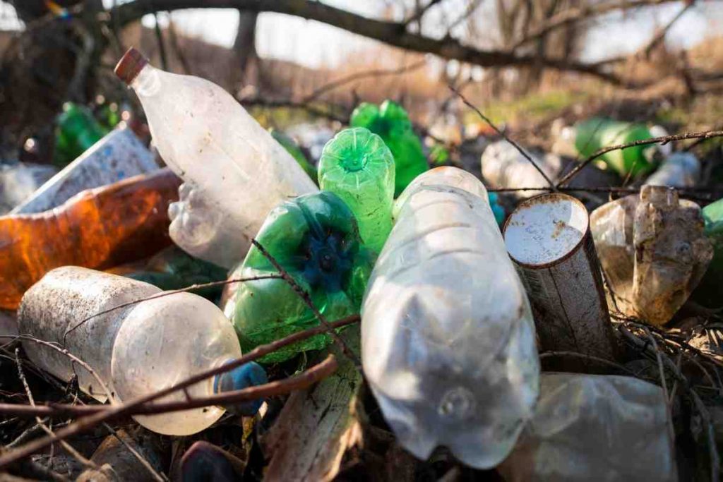
Scoperta una montagna di rifiuti nascosti, si trova in uno dei posti più belli di Italia
In uno dei posti più belli d’Italia spunta una montagna di rifiuti. E’ allarme danno ambientale, che potrebbe ammontare centinaia di migliaia di euro L’Italia …